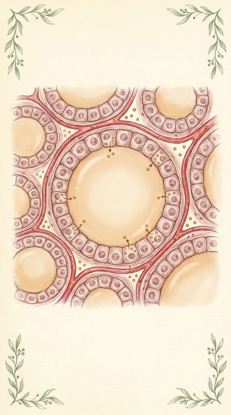
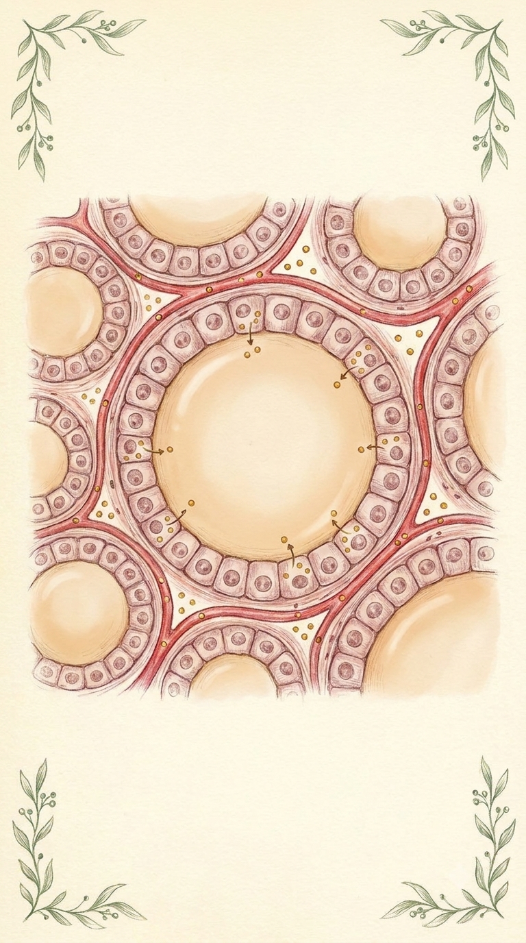
Same hand-drawn medical atlas style as the reference images: colored pencil on cream paper, soft burgundy and rose tones, small green leafy sprigs in the corners, no text, no letters, no labels. Vertical 9:16. Histologically accurate magnified view inside the thyroid gland, like a hand-drawn atlas illustration of thyroid tissue: several round thyroid follicles of different sizes, each a ring of a single layer of cube-shaped follicular cells with round nuclei, filled with smooth pale amber colloid. Thin red capillaries run between the follicles. Small golden-yellow dots (iodine) float in the capillaries next to the follicle walls, some passing into the cells. Gentle warm light, clear readable composition, one large follicle in the center, space at the bottom third for subtitles.